Back Of Neck Anatomy : Posterior Neck muscles | Physical Therapy - Powerlifting ... - The spine runs from the base of your skull down the length of your back, going all the way down to your pelvis.
Back Of Neck Anatomy : Posterior Neck muscles | Physical Therapy - Powerlifting ... - The spine runs from the base of your skull down the length of your back, going all the way down to your pelvis.. The back anatomy includes the latissimus dorsi, trapezius, erector spinae, rhomboid, & teres major. Head and neck trunk and limbs. 3d video tutorials and interactive modules on the anatomy of the back including anatomy of the musculature, vertebral column, joints and ligaments. Surface anatomy and surface markings bibliographic record list of illustrations subject index. The levator scapulae muscle is attached at the top four cervical vertebrae (c1 to c4) and runs down the side of the neck to attach at the top of the shoulder blade (scapula).
Demonstrate a neck and vertebral column; Surface anatomy and surface markings bibliographic record list of illustrations subject index. Apply anatomical knowledge in evaluating movement of the axial skeleton; The structure is, of course, an important part of the conversation. This article describes the anatomy of the head and neck of the human body, including the brain, bones, muscles, blood vessels, nerves, glands, nose, mouth, teeth, tongue, and throat.
Vintage illustration from "The Anatomy Of The Human Head ... from i.pinimg.com The anterior jugular vein (v. Surface anatomy and surface markings bibliographic record list of illustrations subject index. When most people mention their back, what they are actually referring to is their spine. Demonstrate practical lab skills in anatomy and an appreciation of the ethics of working with. This was first described by the sloan kettering group in 19812 and has since been adopted by the american head and neck society (ahns) for the classification of neck dissection with various modifications ( fig. The structure is, of course, an important part of the conversation. The spine runs from the base of your skull down the length of your back, going all the way down to your pelvis. Dummies has always stood for taking on complex concepts and making them easy to understand.
Head and neck trunk and limbs.
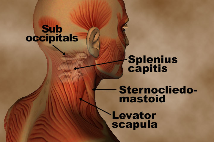
The posterior muscles of the neck are primarily concerned with head movements, like extension. Anterior muscles of the neck. Many in the neck help to stabilize or move the head. The spine runs from the base of your skull down the length of your back, going all the way down to your pelvis. Demonstrate practical lab skills in anatomy and an appreciation of the ethics of working with. The structure is, of course, an important part of the conversation. Jugularis anterior) begins near the. Apply anatomical knowledge in evaluating movement of the axial skeleton; This atlas is a comprehensive and affordable learning tool for residents and medical students and especially for radiologists and surgeons. This entry was posted in anatomy by admin. Anatomy if neck and back diagram. 3d video tutorials and interactive modules on the anatomy of the back including anatomy of the musculature, vertebral column, joints and ligaments. 12 photos of the anatomy of the back of the neck.
They control the scapulae (shoulder blades), which play a role in shrugging, neck movement, head. Some important structures contained in or passing through the neck include the seven cervical vertebrae and enclosed spinal cord, the jugular veins and carotid arteries, part of the esophagus, the larynx. The head rests on the top part of the vertebral column, with the skull joining at c1. The swansea head u0026 neck ultrasound 1899 antique superficial structures of neck surgical anatomy. The neck is the area between the skull base and the clavicles.
Neck Pain - OrthoInfo - AAOS from orthoinfo.aaos.org The swansea head u0026 neck ultrasound 1899 antique superficial structures of neck surgical anatomy. Anterior muscles of the neck. 3d video tutorials and interactive modules on the anatomy of the back including anatomy of the musculature, vertebral column, joints and ligaments. Female neck anatomy anatomy of human neck anatomy human. Despite being a relatively small region, it contains a range of important anatomical features. The neck is the area between the skull base and the clavicles. Anatomists tend to classify the body into during muscle traction, the cheeks are pulled together, which makes food move back and forth. Neck anatomy, lateral neck, central neck, posterior neck, cervical fascia.
Anterior muscles of the neck.
Anatomy of the head and neck: Digastric, mylohyoid, geniohyoid, stylohyoid infrahyoid muscles: « back show on map ». Many in the neck help to stabilize or move the head. The levator scapulae muscle is attached at the top four cervical vertebrae (c1 to c4) and runs down the side of the neck to attach at the top of the shoulder blade (scapula). The posterior muscles of the neck are primarily concerned with head movements, like extension. Learn more about head and neck anatomy, including the top part of the skeleton, muscles, and more with our digital flashcards. It runs down the back part of the neck, and opens into the external jugular vein just below the middle of its course. Choose from 500 different sets of flashcards about neck anatomy back neck upper on quizlet. How to view the anatomical labels. The structure is, of course, an important part of the conversation. Clinically, surface anatomy is used to split the neck into anterior and posterior triangles which provide clues as to the location of specific structures. Your neck is like no other part of the vertebral spinal column and enables your head and neck a wide range of motion.
Learn about these muscles, their locations & functional the traps are quite a complex set of muscles. Your neck is like no other part of the vertebral spinal column and enables your head and neck a wide range of motion. This was first described by the sloan kettering group in 19812 and has since been adopted by the american head and neck society (ahns) for the classification of neck dissection with various modifications ( fig. Clinically, surface anatomy is used to split the neck into anterior and posterior triangles which provide clues as to the location of specific structures. Anatomy if neck and back diagram.
Anatomy and Pathology for bodyworkers - Real Bodywork from www.realbodywork.com Despite being a relatively small region, it contains a range of important anatomical features. We've largely focused on the physical aspect of our spinal anatomy in this series. This article concerning the anatomy of the head and neck area gives you a clear structure at hand to see anatomy and function of the regions of the lower face. Neck, in land vertebrates, the portion of the body joining the head to the shoulders and chest. Digastric, mylohyoid, geniohyoid, stylohyoid infrahyoid muscles: The levator scapulae muscle is attached at the top four cervical vertebrae (c1 to c4) and runs down the side of the neck to attach at the top of the shoulder blade (scapula). This article describes the anatomy of the head and neck of the human body, including the brain, bones, muscles, blood vessels, nerves, glands, nose, mouth, teeth, tongue, and throat. Head and neck anatomy is important when considering pathology affecting the same area.
Jugularis anterior) begins near the.
How to view the anatomical labels. Head and neck trunk and limbs. They control the scapulae (shoulder blades), which play a role in shrugging, neck movement, head. Your neck is like no other part of the vertebral spinal column and enables your head and neck a wide range of motion. This is often a result of incorrect posture. The anterior jugular vein (v. This was first described by the sloan kettering group in 19812 and has since been adopted by the american head and neck society (ahns) for the classification of neck dissection with various modifications ( fig. Jugularis anterior) begins near the. Posterior triangle of the neck boundari… pretracheal fascia b. The structure is, of course, an important part of the conversation. It runs down the back part of the neck, and opens into the external jugular vein just below the middle of its course. 12 photos of the anatomy of the back of the neck. Choose from 500 different sets of flashcards about neck anatomy back neck upper on quizlet.


0 Komentar